A visual field test, also called a field of vision test, vision field test, or eye field test, is essential for diagnosing and monitoring glaucoma, neurological disease, retinal pathology, and functional driving vision.
But as every eye doctor and technician knows, even the best perimeter can’t fix poor testing technique. Unreliable results, high false positives, fixation losses, and anxious patients all reduce the clinical value of your visual field testing.
This guide explains how to get better visual field test results by focusing on:
- Technician training and confidence
- Patient preparation and reducing visual field test anxiety
- Optimizing the test environment
- Active monitoring during the exam
- Knowing when to repeat or discard results
- Matching the pattern (24-2, 10-2, C-40, Esterman, Superior 36, etc.) to the clinical need
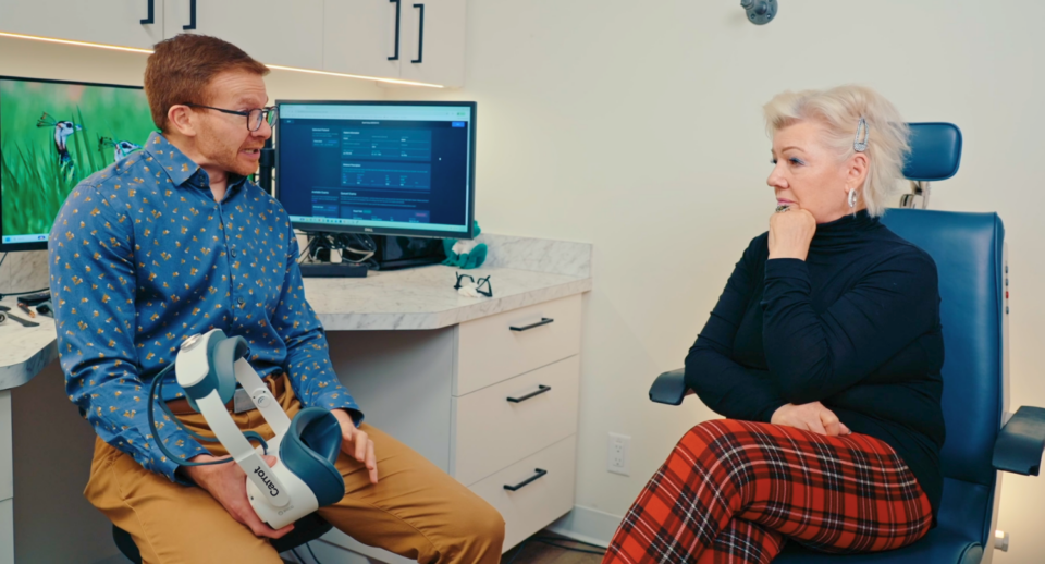
Throughout, we’ll highlight how the Carrot Visual Diagnostics Platform helps practices standardize workflows and improve the quality of visual field test results across your patient base.
1. Train Technicians for Accurate Visual Field Testing
Reliable visual field testing starts with the examiners. Techs need more than button-press skills. They need to understand:
- What makes a good vs. a bad visual field test
- How false positives, fixation losses, and false negatives appear on the report
- When to pause, coach, or restart a test
- How to respond when patients say, “I hate visual field tests”
With Carrot, practices can support technician training through:
- A detailed online user manual and quick-start guides
- Training videos and resources like:
- Unlimited onboarding sessions with a dedicated customer success manager
- Live guidance and troubleshooting support for real-world cases
The result: technicians feel confident, providers trust the data, and your practice is recognized as a leader in virtual visual field testing.

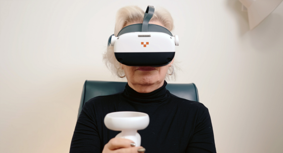
2. Prepare Patients Before the Test (and Reduce Visual Field Test Anxiety)
Many common issues, such as false positives, poor fixation, and test fatigue, begin before the test begins. Good preparation is one of the best tips for taking a visual field test.
Use a Standardized Script
Give every patient the same, clear explanation:
- What the visual field test is
- That there’s no pass/fail, only measurement
- That it’s normal not to see every light
- That they should respond only when they are sure they see a stimulus
This directly addresses common patient concerns, such as how to pass a visual field test (or how to fail one). You’re reframing it as cooperation and accuracy, not performance.
Screen for Fatigue, Pain, and Stress
Before testing:
- Ask about headaches, fatigue, and pain
- Offer restroom or breaks first, not mid-test
- Consider splitting testing across visits for fragile or cognitively limited patients
Clarify Expectations
Patients frequently ask their doctors, can a visual field test be wrong? Assuage their concerns by letting them know:
- You’re measuring how their vision behaves today, not grading them
- It’s okay if the first test is a “warm-up” and needs to be repeated
This alone can reduce anxiety about visual field testing and improve reliability.
3. Set the Stage: Environment, Comfort, and Equipment Setup
The test environment is a powerful lever for improving visual field test results.
Control Lighting
Even with VR-based platforms like Carrot, a dim and quiet room:
- Reduces distraction
- Lowers anxiety
- Improves fixation
Support Multilingual Instructions
For many patients, language barriers are the most significant source of confusion. The Carrot headset offers multilingual audio instructions (41 languages).
Evaluate Ocular and Eyelid Factors
Before starting the exam:
- Check for dry eye and instill tears if needed
- Assess for ptosis; if the lid obstructs the superior field, tape the eyelid per your protocol
- Use Superior 36 or Superior 64 patterns when evaluating ptosis or blepharoplasty cases, and link to:
Ensure Proper Positioning and Alignment
Whether using a tabletop perimeter or a Carrot headset:
- Ensure correct working distance and fixation alignment
- If using trial lenses, position them carefully to avoid rim artifact
- With VR headsets, center the headset to avoid generalized depression or localized artifacts
Small improvements here dramatically reduce complaints like failing the visual field test due to poor setup.
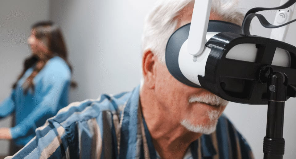
4. Conduct the Test with Active Monitoring
The worst thing a technician can do is press start and walk away. Active monitoring is critical for catching issues early:
- Watch the fixation tracker and eye-tracking indicators
- Look for excessive false positives (“trigger-happy” patients)
- Watch for eye closure, drowsiness, or confusion
- Pause and rest if performance declines noticeably
- Restart when you see a clearly unusable early pattern
With Carrot, techs can see real-time feedback and auto-pausing for inactivity, making it easier to intervene before an unusable result is produced.

5. Know When to Discard or Repeat a Visual Field Test
Not every test should remain in the chart forever.
Reliability Flags to Watch
- False Positives > 15–20%
- Often related to anxiety or over-anticipation
- Can create artificially “good” fields that don’t match reality
- Fixation Losses
- A few may be acceptable, especially in advanced disease
- Consistent, high fixation losses + inconsistent fields over time = retest
- False Negatives
- Common in advanced glaucoma or neurological disease
- Evaluate in context of pattern, prior exams, and structural findings
- Internal Consistency
- Compare today’s visual field to prior results
- Ask: does this fit the patient’s clinical picture and OCT / fundus findings?
If a test is clearly unusable, repeat it the same day when possible, once you’ve addressed the root cause.
6. Match the Visual Field Pattern to the Clinical Question
Choosing the correct testing pattern is one of the most common steps that practices miss when wondering how to get better visual field test results. A misaligned pattern often looks “normal” while missing the relevant disease.
Recommended Patterns for Common Clinical Scenarios
With Carrot, these patterns are all available from the same headset, making it easy to match testing to clinical indications instead of forcing one pattern to answer every question.
| Exam Pattern | Use Case | Related Resource |
|---|---|---|
| Central 24-2 & 30-2 | Glaucoma, optic neuropathy, general screening of the posterior pole | |
| Central 24-2C | Macular-involving glaucoma, central field loss, dense testing near fixation | |
| Central 10-2 | Plaquenil (HCQ) toxicity, macular disease, advanced glaucoma | |
| C-40 & N-30 | Fast visual field screening for general or initial evaluations | |
| Superior 36 / Superior 64 | Ptosis, dermatochalasis, taped vs. untaped eyelid testing for eyelid surgery and insurance approval | |
| Esterman / Full Field 120 | Driving standards, licensing, functional binocular field, peripheral vision monitoring | |
| Kinetic Visual Field | Peripheral field mapping, neurological disease, complex or unusual defects |
7. Interpreting and Tracking Visual Field Test Results Over Time
Better visual field test results empower eye doctors to interpret and trend them.
Look Beyond a Single Field
- Use progression reports and trend analysis
- Compare to OCT (RNFL, GCC), fundus imaging, and IOP
- Re-baseline when switching from tabletop to virtual devices
Final Thoughts
Improving visual field test quality is equal parts finding the best virtual visual field device for your practice and building repeatable, technician-friendly workflows. By standardizing patient instructions, optimizing the testing environment, actively monitoring exams, and choosing the right patterns, you can dramatically help you get better visual field test results.
With Carrot, practices can:
- Reduce variability
- Test more comfortably and efficiently
- Document reliable results across diverse patient populations
That’s how you transform “I hate visual field tests” into smoother, more accurate experiences for both patients and providers.