What Is Confrontation Visual Field Testing?
A confrontation visual field test is one of the simplest and quickest ways to assess a person’s visual field, the total area in which objects can be seen in peripheral vision while the eye is focused on a central point. This test is often part of a standard eye exam or neuro-optometric screening and helps detect early signs of visual field loss caused by conditions such as glaucoma, stroke, optic neuritis, or pituitary tumors.
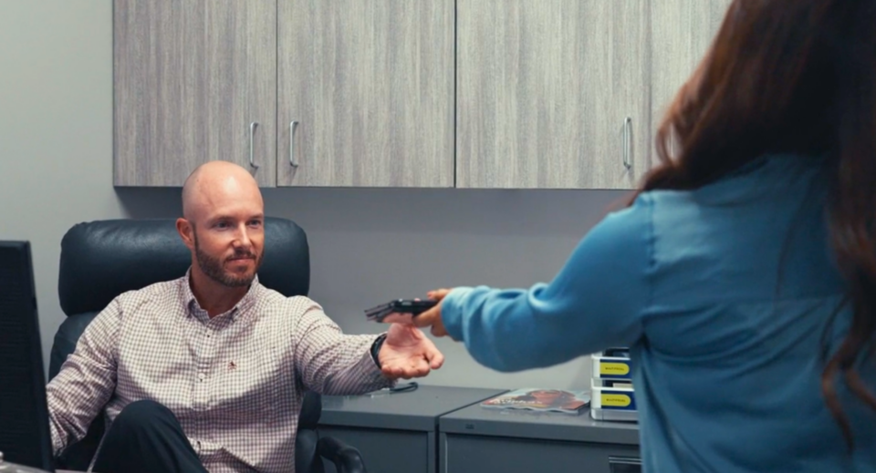
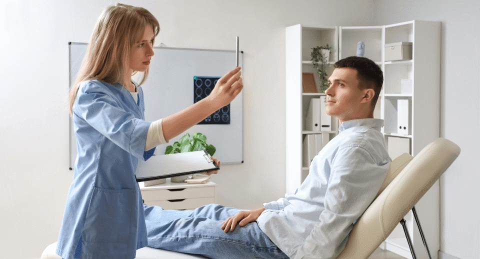
During the confrontation visual field test, the patient covers one eye while focusing on the examiner’s face. The clinician then moves a small target, like a finger or pen tip, into different parts of the patient’s visual field. The patient signals when they can see the object, allowing the examiner to map areas of reduced or lost peripheral vision.
While the confrontation visual field test is not as sensitive as automated perimetry with a virtual visual field device (or the Humphrey visual field test), it remains an essential first-line visual field screening test, particularly in primary care or emergency settings where quick functional assessment is needed.
Common reasons for performing a confrontation visual field test include:
- Glaucoma visual field loss detection
- Visual field testing for ptosis or blepharoplasty
- Neurological visual field defects, such as hemianopia or quadrantanopia
- Driving visual field test screening in patients reporting peripheral vision concerns
Because the visual field defect patterns can point to specific eye or brain disorders, confrontation testing often serves as the gateway to more advanced perimetry testing or virtual visual field testing.
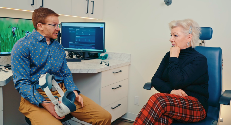
How Confrontation Testing Compares to Virtual Visual Field Testing with Carrot
Traditional confrontation testing is subjective. It relies heavily on patient cooperation and examiner consistency. While useful for gross defects, it lacks the precision to quantify visual field defects or monitor subtle progression in diseases like glaucoma.
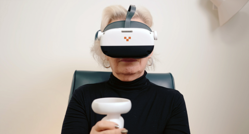
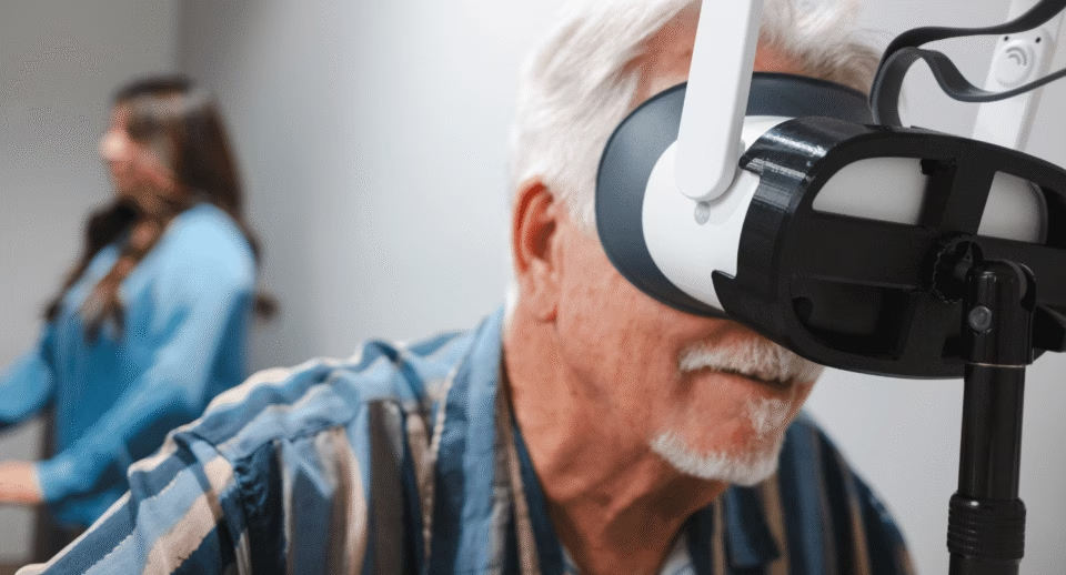
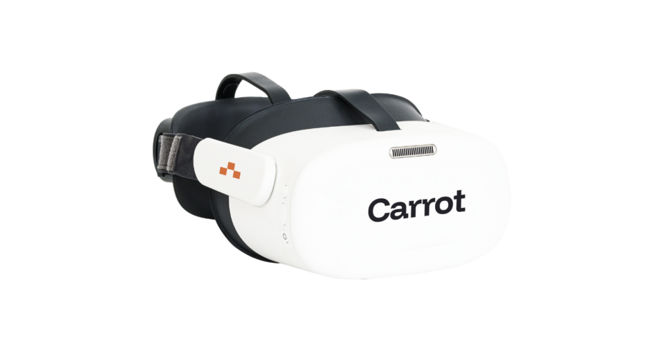
That’s where Carrot’s virtual visual field testing offers more significant diagnostic capabilities. Using virtual reality (VR)-based perimetry, Carrot combines accuracy, portability, and patient comfort into a single device. Instead of a static bowl perimeter or handheld target, patients wear a VR headset that presents controlled visual stimuli while tracking eye fixation and responses in real time.
| Feature | Confrontation Visual Field Test | Virtual Field (VR Visual Field Test) |
|---|---|---|
| Accuracy | Detects large field defects only | Detects subtle scotomas and early glaucoma changes |
| Quantification | Qualitative | Quantitative with detailed visual field maps |
| Equipment | None required | Lightweight portable headset |
| Environment | Requires a well-lit room and a trained examiner | Can be performed anywhere with minimal setup |
| Patient Comfort | Quick and simple | Immersive, more engaging experience |
| Tracking | Manual | Automated eye tracking and response capture |
Carrot also supports tests equivalent to Humphrey 24-2, 10-2, and Esterman visual field protocols. The VR platform enables automated perimetry testing and progression monitoring, ideal for glaucoma management, postoperative recovery, or patients with mobility challenges.
By combining automated visual field analysis with cloud-based reporting, Carrot provides clinicians with accurate visual field test results comparable to those from established devices such as the Humphrey Field Analyzer or Goldmann perimetry, but in a more accessible, patient-friendly format.
Best Practices for Confrontation Visual Field Testing
Although simple, confrontation testing should be performed with care to ensure accuracy. Below are key best practices to optimize results and minimize variability:
1. Standardize the Testing Environment
Perform the test in a well-lit room with a non-distracting background. The patient should sit at eye level with the examiner, about an arm’s length away. Inconsistent lighting or distractions can lead to false negatives, particularly when testing for peripheral vision loss or visual field defects in glaucoma.
2. Ensure Proper Patient Fixation
Ask the patient to fixate steadily on your open eye or the bridge of your nose. Loss of fixation can lead to inaccurate mapping of the visual fields, especially for central or paracentral visual field defects. Reinforce the importance of maintaining gaze throughout the test.
3. Use Both Static and Kinetic Methods
- Static confrontation: Present your fingers in different quadrants and ask the patient to identify which are moving.
- Kinetic confrontation: Move a target slowly from the periphery toward the center. This dual approach increases sensitivity for detecting subtle visual field loss, especially near the midline.

4. Compare Bilateral Responses
Always compare the right and left eyes. Asymmetric or unilateral responses may indicate optic nerve or retrochiasmal lesions. Comparing both eyes helps differentiate normal variations from true defects.
5. Document Clearly
Record which quadrants showed reduced or absent responses. Use consistent terminology (e.g., “inferior nasal field defect” or “superior temporal scotoma”). Proper documentation ensures continuity if the patient later undergoes automated perimetry or virtual visual field testing.
6. Refer for Automated or Virtual Testing When Needed
If the confrontation test reveals any abnormal visual field test results, refer the patient for comprehensive perimetry testing, such as Humphrey visual field testing, Goldmann perimetry, or Carrot’s virtual visual field test.
Carrot’s portable visual field testing equipment enables clinicians to quickly and accurately validate confrontation findings, even outside traditional clinic settings.
Final Thoughts
The confrontation visual field test remains a valuable, low-tech screening tool for detecting gross visual field defects, but it shouldn’t stand alone. Advances in virtual visual field technology, like those pioneered by Carrot, offer a more consistent, quantifiable, and accessible way to monitor visual health across diverse patient populations.
Integrating both approaches, starting with confrontation testing and confirming with automated VR perimetry, provides the best of both worlds: clinical efficiency and diagnostic precision.
Whether screening for glaucoma visual field loss, assessing ptosis visual field impact, or documenting visual field results for blepharoplasty, adopting these best practices ensures patients receive the most accurate, evidence-based care possible.