A Comprehensive Guide for Early AMD Screening
The Amsler grid test is impressively simple and ideal for home monitoring of macular degeneration. A 20 x 20 grid with a singular central spot, this early AMD screening tool can help identify changes in central vision caused by macular degeneration or visual pathway abnormalities — even when symptoms are subtle.
Amsler Grid Overview
Developed by Swiss ophthalmologist Marc Amsler in the 1940s, the Amsler grid is a square chart of horizontal and vertical lines with a central fixation point. It tests the integrity of the macula and the surrounding central retina, as it’s particularly sensitive to disruptions in the retinal pigment epithelium or photoreceptor alignment.
To conduct the exam, ask patients to look at the central dot, one eye at a time. They should report any irregularities in the surrounding lines, like waviness, blank spots, or areas that appear dim or blurry. From there, you can continue testing for macular degeneration or other macular conditions.
Academic references and clinical validation
|
|
The Amsler grid has a sensitivity of 62.7% and a specificity of 68.4%. Despite these gaps in testing ability, it remains one of the most well-known and easiest-to-administer screening tests today. |
|
|
The Amsler grid layout is similar to Gestalt optical illusions, and recent research suggests that this test triggers a “filling in” phenomenon, which can lead to false negatives. |
|
|
A recent study highlighted that although the Amsler grid is accessible and easy to use, its sensitivity is low and nonspecific, making further testing mandatory. |
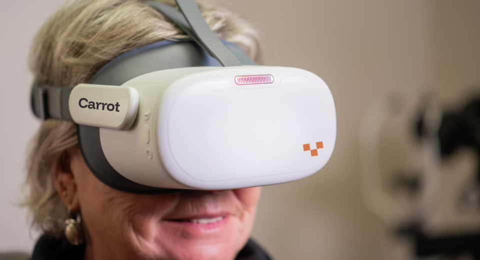
Carrot is proud to be the most trusted provider of virtual visual field testing in the United States and Canada.


The Amsler Test at a Glance
For patients without macular conditions, Amsler grid lines appear straight and uninterrupted. When the macula is damaged, some areas appear warped, missing, wavy, blurry, or even discolored. AMD-related structural changes, such as drusen deposits, distort the way light is detected and processed in the central retina. This information reveals physiological blind spots within the central 10 degrees, but provides no insight into peripheral vision. Eye care providers appreciate how quickly and easily they can get a general sense of AMD progression. Still, the rate of false negatives and lack of specificity can leave many questions unanswered.
Patients might notice a range of aberrations, such as the following:
- Metamorphopsia: Lines appear bent, wavy, or curved
- Scotomas: Missing or darkened areas in the grid
- Micropsia or macropsia: Lines appear squashed or spread apart
- Blurring: Areas that appear foggy or smudged
Patients who take this test at home should mark areas of concern directly on a printed grid and bring the results to their appointments for your evaluation.
Pros and Cons of Amsler Grid Testing
The pros and cons that follow can help guide you toward the ideal scenarios to incorporate this test into your patients’ diagnostic assessments.
|
Pros
|
Cons
|

List of Ocular Diseases Monitored and Diagnoses Identified by Amsler Testing
|
|
Example Amsler Grid


Billing and Coding for Amsler Tests
This test usually fits into an extended visual field exam, which is billable using CPT code 92083, or a routine screening (CPT codes 92004 and 92014). The Amsler grid test isn’t separately billable. Reimbursement varies greatly and depends on your practice’s location, setting, and the precise type of exam you’re providing.
When is the Amsler test required?
Patients diagnosed with, or at risk for, macular degeneration, central serous retinopathy, macular pucker, diabetic macular edema, retinal diseases, or macular injury will need this test. It’s often recommended that patients with known AMD use this test daily to monitor their condition.
Is Amsler testing required for driver’s licenses?
Patients with macular degeneration can often continue driving, even with blind spots and visual disturbances. In the United States, each state sets a minimum for visual acuity, and many also require visual field perception. Esterman exam results are usually sufficient.
Complete Your Comprehensive Exams with Carrot
Consider the Amsler Grid just one component of a comprehensive eye exam. Eye care professionals will also need to interpret the patient’s experience, conduct further testing, and make an appropriate treatment plan.
Carrot’s functional vision and visual field tests can provide more insight and help monitor disease progression. You can add Carrot to your testing routines to deliver comprehensive, precise, and patient-friendly eye exam experiences at scale.
Want all 23 of our comprehensive test guides in your inbox? Fill out the form below.

Frequently Asked Questions
Generally no. Most carriers bundle it into the eye exam. Unlisted code 92700 can be attempted but is rarely paid.
Patients report wavy lines (metamorphopsia) or small central scotomas often preceding OCT-visible fluid by weeks.
Daily or at least twice weekly self-monitoring is advised.
Appearance of a new distortion ≥ 1 grid square or enlargement of an existing scotoma warrants same-week retinal evaluation.
Carrot is always enhancing its solution. We are striving to add valuable tests to our offering as soon as we can validate them to meet our high-quality standards and those of our provider customers.