In a busy clinic, you likely follow a set checklist. You are familiar with the components of a thorough screening and know how to follow up with established patients. The most comprehensive eye exams prioritize tests such as acuity, refraction, retinal imaging, and intraocular pressure. These are core components, but thorough eye exams should always include visual field testing. This way, you can measure functional vision, catch progressive conditions sooner, and provide more complete patient eye care.
The Role of Visual Field Testing in Eye Care
A visual field test evaluates the full horizontal and vertical range of a patient’s peripheral vision. It maps both the sensitivity and extent of vision that no acuity chart can measure. Visual field testing detects functional deficits, not just structural abnormalities, and can specify where patients may be experiencing scotomas.
There are many types of visual field tests, but for general eye care, there are two most useful categories:
- Automated static perimetry tests fixed points at varying intensities. This testing modality is ideal for tracking glaucoma and other progressive diseases because it clearly and consistently maps blind spots and changes in the visual field.
- Kinetic perimetry (also called Goldmann perimetry) uses moving stimuli to map the boundaries of the patient’s visual field. This technique is more sensitive to neurological defects.
Clinical Justifications for Standardization
Visual field tests reveal functional changes that other modalities often miss. Beyond the clinical rationale, incorporating visual field testing into every comprehensive eye exam routine strengthens your practice in practical ways. As part of the complete eye care process, visual field testing can improve outcomes.
Early Detection of Ocular Diseases
Glaucoma is often asymptomatic in its early stages. Patients often don’t notice symptoms until they are advanced, and even in the later stages, they often maintain good acuity. Functional losses in the visual field often precede visible structural damage, and early detection of glaucoma can help preserve vision for longer.
The diagnostic capabilities of visual field tests also apply to early retinal pathology and optic nerve disorders. Visual field testing is one of the most essential tools for measuring optic nerve health. This way, you can reveal neuropathies or retinal damage well before they become apparent on imaging.
️ Read this case study to learn how Scott and Christie Eyecare Associates ensure Glaucoma patient treatment plan compliance with Carrot.
Neurological Disorders
Brain tumors, strokes, multiple sclerosis, and other neurological events may manifest through field of vision changes. In fact, visual field changes are often the first signs of these serious conditions.
Routine field testing gives you a functional screen for neurological conditions so that visual field data can guide timely referrals to neurology, oncology, internal medicine, or oculoplastics. Integrating medical care into your optical shop is another opportunity to preserve sight and intervene earlier.
️ Pupillometry is also highly sensitive for detecting neurological disorders.
Monitoring Disease Progression
Consistent visual field testing provides an opportunity to identify subtle changes, initiate targeted treatments when necessary, and accurately document disease progression.
At Carrot, we believe in the power of progression analysis so much that we designed our solution to automatically generate a progression analysis report after just two tests on the same eye, so you can quickly track quantitative changes from the baseline.
Managing progressive conditions like glaucoma, tracking medication toxicity, and monitoring the success of pre- and post-surgicalcataract patients are possible when visual field testing is a routine part of the exam process.

Business and Patient Care Benefits
Visual field testing adds clarity. More data means more informed decisions, which leads to more targeted care. Incorporating visual field testing into your comprehensive eye exams gives you objective evidence to support diagnosis, monitor treatment efficacy, and justify interventions.
Patients also appreciate thoroughness. Integrating visual field testing can distinguish your practice, improve trust, and even serve as a marketing point for high-quality care.
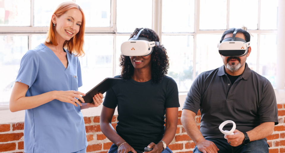
Some patients find traditional tabletop perimetry awkward or uncomfortable, but using Carrot’s lightweight headset is much more patient-friendly. And because the Carrot headset is portable, you don’t need a dedicated workspace, unlike traditional methods.
️Read this case study to learn what made Nicole Hattaway, the Assistant Practice Administrator for the Georgia Center for Sight, say:
“In our main office, we have a dedicated testing area, but not in our satellite offices. With a Pro Carrot subscription, we can conduct the test in any exam room. It’s installed on all our computers. We can bring the field test to the patient instead of the other way around.”
From a medicolegal standpoint, documenting visual field status reduces liability. Managing glaucoma and responding to visual complaints will require a functional baseline. Regular visual field testing strengthens documentation and helps you demonstrate your standard of care over time.
With Carrot, visual field exams are far more accessible and time-efficient. Traditional tabletop perimeters typically take 5 to 7 minutes per eye, but Carrot cuts that time by over 2 minutes per eye and easily integrates into the exam lane or pretest workflow. Conduct tests anywhere patients feel comfortable.
Rethinking Comprehensive Eye Care
If you’ve been skipping visual field testing as part of your complete eye exams, you’re not seeing the whole picture of patient eye health. Visual field testing could reveal glaucoma, optic neuropathies, retinal conditions, macular degeneration, and many other conditions long before they become symptomatic. Following up on the progress of these conditions is only possible with a baseline, and incorporating visual field testing into your workflow doesn’t have to be a burden. It’s part of a functional vision assessment, and with Carrot, it’s faster, more accessible, and patient-friendly.