A Comprehensive Wide-Field Glaucoma and Lesion Assessment Guide
Patients usually don’t notice their peripheral vision until it’s lost or reduced. At that point, they may realize they struggle with spatial orientation or miss hazards that fall outside their line of vision. They may have normal central visual acuity, but significant loss at the periphery. The 60-4 peripheral visual field test measures these changes, especially when they’re related to neurological conditions.
The 60-4 peripheral visual field test helps clinicians map out functional vision loss in areas beyond the central 30 degrees, making it critical for neurological conditions and advanced glaucoma.
This specialty threshold test isn’t usually part of routine exams, and not all perimeters can conduct the 60-4 peripheral visual field test. While not currently supported by Virtual Field, the 60-4 is an important wide-field glaucoma test and valuable for chiasmal lesion assessment and advanced disease tracking.
What Is the 60-4 Peripheral Visual Field Test?
If you have a Humphrey Field Analyzer, you can conduct the 60-4 peripheral visual field test using a suprathreshold or threshold protocol. Unlike the more standard 24-2 or 30-2 exams, which target the central macula and surrounding areas, the 60-4 peripheral visual field test presents stimuli at points extending much farther into the peripheral retina. This ability allows you to assess the 60-4 peripheral visual field, which is essential for detecting neurological conditions and supporting chiasmal lesion assessment in cases of visual pathway compression or damage.
This test measures visual sensitivity up to 60° in all directions. Unlike most threshold exams, which focus on the central 30°, this grid captures functional deficits in the outermost regions of the visual field.
The 60-4 peripheral visual field test is used selectively, usually only when peripheral pathology is suspected, such as neurological conditions, retinal degenerations, and cases where central vision remains normal but the patient complains of spatial or mobility issues. In the absence of this test, a portable full-field perimeter may offer limited but useful insight into peripheral visual awareness.
Academic references and clinical validation
| | Extended peripheral visual field testing is surprisingly controversial. An older article in Healio asserts that the 60-4 is rarely necessary, while more recent conversation explores its clinical and algorithmic applications. |
| | Recent research has shown that AI can accurately predict 60-4 visual field defects in healthy subjects based on 3D facial reconstruction. This pilot study suggests that there is room for AI tools to improve the interpretation of peripheral field results by accounting for individual facial features. |
| | The 60-4 is not without its limitations. A study found that healthy males had more peripheral 60-4 visual field defects than females, likely due to differences in facial structure. This insight further suggests that non-clinical factors can impact this far-peripheral measurement. |
These findings reinforce the clinical importance of the 60-4 peripheral visual field test for comprehensive assessment of retinal and neurological function, especially when paired with newer portable full-field perimeter technologies.
Did You Know?
The average Carrot customer gets an extra 62 working hours back thanks to the workflow efficiencies from our Visual Diagnostics Platform.


Wide Field Glaucoma Test: How the 60-4 Peripheral Visual Field Test Works
This test measures extreme peripheral sensitivity beyond the 30° radius. The 60-4 peripheral visual field test is almost exclusively conducted on the Humphrey Field Analyzer in either full or suprathreshold mode. It’s best used in patients with suspected peripheral field loss due to neurological, hereditary, or vascular diseases. Because it’s used for wide field glaucoma testing, the 60-4 offers value where traditional 24-2 or 30-2 tests may miss peripheral damage.
Conducting the 60-4 peripheral visual field test similarly to other perimetry exams, you or your technician will ask the patient to focus on a central point and respond to stimuli in the far periphery. For practices that conduct frequent visual field exams, this shouldn’t be an unusual process. However, there could be false positives due to factors such as facial structure, patient understanding, people with narrow pupils, and others. It’s also a relatively rare test, which can make interpretation a challenge.
In clinics without access to this exam, consider using a portable full-field perimeter to facilitate peripheral field analysis, especially for patients with mobility concerns.
Benefits and Drawbacks of the 60-4 Wide Field Vision Test
The pros and cons that follow can help guide you toward the ideal scenarios to incorporate this test into your patients’ diagnostic assessments.
Pros
| Cons
|

Conditions Detected with 60-4 Peripheral Testing and Chiasmal Lesion Assessment
The 60-4 is particularly useful for wide field glaucoma testing and diagnosing advanced or uncommon visual conditions. It’s also an effective chiasmal lesion assessment tool when patients present with bitemporal hemianopsia or post-surgical changes.
|
|

CPT Code, Billing, and Reimbursement for 60-4 Visual Field Exams
CPT code 92083 is used for extended perimetry exams such as the 60-4 peripheral visual field test, often categorized under wide field glaucoma tests. The CMS physician fee schedule reimbursement can be anywhere from $25 to $80 per exam, but this amount will depend on your practice’s location, setting, and the precise type of exam you’re providing.
When is the 60-4 visual field test required?
The 60-4 is reserved for particular cases. Patients with advanced glaucoma, retinitis pigmentosa, tumors, and certain neurological conditions should have this exam about once a year. If this test is already part of your workflow, consider it for patients who complain of changes in their peripheral vision or trouble noticing hazards at the edge of their vision.
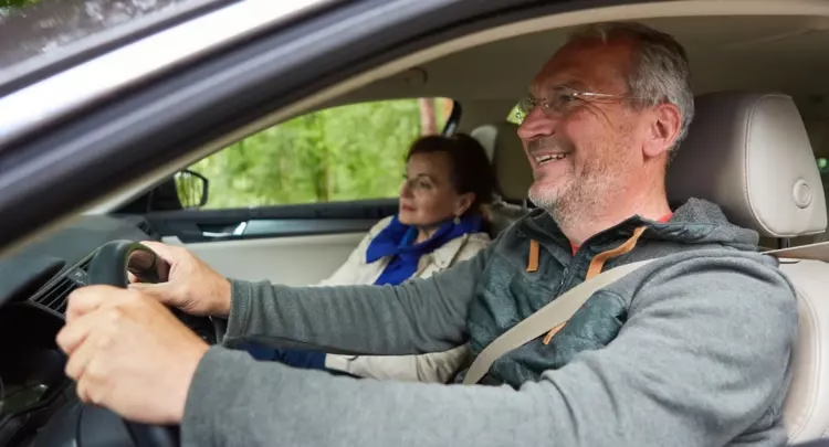
Is the 60-4 required for driver’s licenses?
Generally, no. However, in the state of Virginia, to determine visual field loss, the DMV requires the results of a visual field test that measures the central 24 to 30 degrees of the visual field, and testing needs to be completed to 120 degrees (60 degrees from the point of fixation), therefore a 60-4 or equivalent test is acceptable.
Start Conducting Comprehensive Vision Exams with Carrot
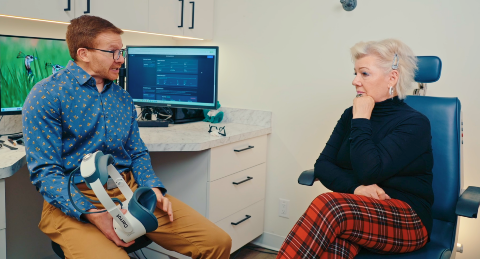
If your practice uses all the tests available on your tabletop perimeter, consider a portable full-field perimeter like Carrot. When you need faster, more comfortable, more patient-friendly eye exams, Carrot’s portable headset fits seamlessly into your practice. Patients with mobility issues may not be able to sit at the tabletop perimeter for long periods, and will find the Carrot headset much more accessible. Others might benefit from Carrot’s 41 audio language options to help guide the testing process. Compare Virtual Field models, plans, and test suites to match your practice’s unique needs.
Want all 23 exam guides in one place?
Download our comprehensive guide for 160+ pages of insights.

Frequently Asked Questions
Choose 60-4 for pituitary adenomas, optic-tract lesions, late-stage glaucoma, or any condition that may cause peripheral constriction beyond 30°.
The 60-4 test grid samples threshold points out to 60 degrees eccentricity in all meridians, doubling central point spacing to keep test time reasonable.
Carrot is always enhancing its solution. We are striving to add valuable tests to our offering as soon as we can validate them to meet our high-quality standards and those of our provider customers.
Bill CPT code 92083 for 60-4 tests. The code applies to any automated threshold pattern, including the 60-4.
Patients accustomed to 24-2 adapt quickly; fixation losses typically normalize after one practice eye, so no separate training visit is needed.
Yes. You can trend Mean Deviation (MD) over time using 60-4 visual field tests.